Prehospital scene management

scene

As hospital doctors working in acute care, we have a considerable amount of control over the scene in which we work. Our ED resus bays have adequate space, lighting, and equipment (which is in the same place every time we need it). We have a huge number of team members we can draw upon for support in our patient care, and with prehospital notification of impending patient arrival we can assemble an appropriate team, set up relevant equipment ahead of time, and establish control over the scene before the patient arrives. We even have waiting areas for family and friends of critically ill patients and can delegate staff to look after them while a resuscitation is occurring.

In the prehospital setting, many of the factors above are unachievable, and to doctors this represents both a source of challenge and considerable discomfort.

One of the most interesting aspects of working as a doctor in the prehospital setting (both in practice and simulation) has been watching my paramedic colleagues in action at a prehospital scene – in particular the skill, calm, and aplomb with which they assess and manage a prehospital scene, and the adaptability with which this process occurs under highly variable circumstances.

While as HEMS doctors it would be uncommon for us to be in a position where we have a significant role in scene management – this role would usually be performed by ambulance staff already at the scene or by the helicopter paramedic – it is important for us to understand the process.

There is comparatively little literature available in this area. There are resources detailing ASSESSMENT of a scene, such as this chapter from the Prehospital Trauma Life Support manual.

With regards to MANAGEMENT of a prehospital scene, the authors of this study, published in EMJ in 2009, conducted interviews with experienced paramedics to generate a theory as to how paramedics manage a scene. The model that resulted was called “the space control theory of paramedic scene management”, which states that paramedics manage a scene by controlling the activities that occur in the space immediately around the patient “Space” is interpreted to include both physical and human (non-physical) elements.

“Although there are physical realities that present problems for scene management, for the most part the management of the scene involves how paramedics interact with other people. Indeed, it is through working with others that paramedics are able to solve the problems presented by both physical and human elements. This means that scene management is a dynamic social activity comprised of social processes.”

This figure from the paper provides overview of the theory:

space control theory

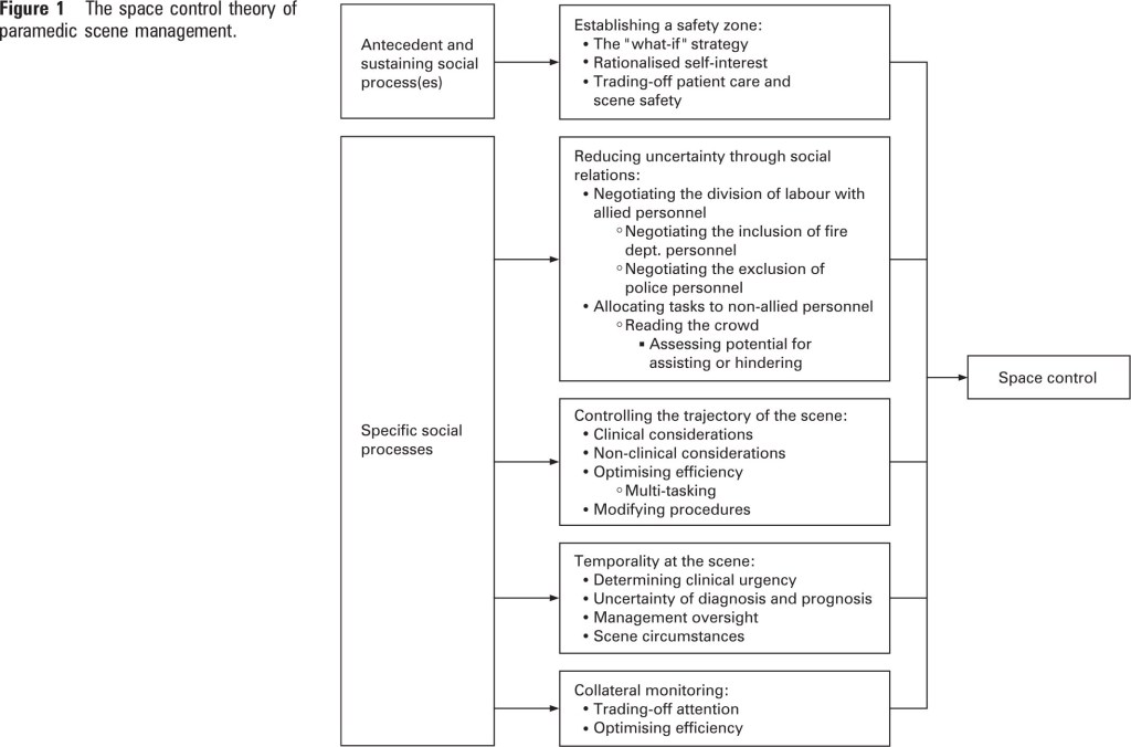
This model has multiple “human factors” elements – analogous to the increasingly recognised importance of human factors in hospital care.

Another useful resource for doctors at a prehospital scene is this 2007 slide set from Tony Smith – ADHB Intensivist, Medical Advisor to St John Ambulance, and Auckland HEMS doctor:

Slide1

Full-text pdf for this post is available here (secure area limited to ADHB staff only – ADHB has online subscription access to this journal through the Philson Library at the University of Auckland School of Medicine)

NAP4 and its implications for prehospital airway management

In 2011 the U.K. Royal College of Anaesthetists and The Difficult Airway Society released a report called NAP4 – the 4th national audit of major complications of airway management.

Full text of NAP4 report

Full text of NAP4 report

The report covered airway complications that occurred in anaesthesia, ICU, and ED settings (approximately 20000 in total). Every reported complication of airway management was analysed for causes and learning points.

The findings relating to ED complications have direct implications for prehospital airway management.

‘Take-home’ messages relating to ED airway management:

  • in the event of an airway complication (most commonly failed RSI), patients were more likely to die in ED or ICU than OR
  • at-risk patients were often not identified prior to the attempt at airway management
  • waveform quantitative capnography should be the standard of care for EVERY intubation
  • situations where the capnography reading was zero (indicating misplaced or completely obstructed ETT) were incorrectly attributed to cardiac arrest (CPR always generates SOME CO2)
  • complications arose when there was a ‘failure to plan for failure’
  • obesity was a major risk factor for airway complications

and, most importantly:

  • in the event of a surgical airway being needed, surgical cricothyroidotomy was almost universally successful, while needle cricothyroidotomy had a failure rate of up to 60%
  • the success of surgical cricothyroidotomy included those where ED doctors (not surgeons) were the ones performing the procedure

Here is an excellent podcast – it is an interview by Cliff Reid of Jonathan Benger, a Professor of Emergency Medicine and one of the authors of the NAP 4 study (sourced from emcrit.org) regarding the implications of NAP4 for emergency department airway management.

So what are the implications for our HEMS service?

The most relevant findings for us form NAP4 are the findings relating to airway complications in ED, more so than anaesthesia or ICU. Patients who we would intubate pre-hospital are those who, if prehospital intubation were not available, would be intubated shortly after arrival in ED. The majority of our doctors are ED-trained, and are most familiar with ED airway management (translation: simple, fast, relatively low-tech, with the fairly standardised approach for the majority of our patients)

Bringing ED airway management to the prehospital arena has its challenges. The patients are more undifferentiated, comparatively under-resuscitated, and there may not have been enough time to get a sense of their ‘trajectory’.Environmental  factors (light, weather, physical access to patient) will have a huge impact on the execution of airway intervention.  We have a lot less equipment – no Glidescope, less rescue devices, and no telephone to call for an anaesthetist and a tech with a trolley full of difficult airway equipment. We may have team members (relatively junior ambulance staff, for example) who have much less experience with RSI than ED nurses who are often part of our RSI team.

Doing the basics right therefore becomes even MORE important:

  • equipment must be effective, functional, and familiar to us through training
  • there must be a ‘shared mental model’ – including a plan for success and a plan for failure – which must be vocalised for every patient with all team members understanding their role
  • we must actively consider patient specific elements that will affect the plan for success and the plan for failure (anatomy, injury, obesity etc)
  • we must be as prepared as possible – if the situation allows, taking several extra minutes to optimise positioning, place nasal cannulae for apnoeic ventilation etc may be crucial
  • we can overcome the disorienting effect of unfamiliar/unfriendly environments by using our RSI checklist – this was we are unlikely to forget something crucial (like capnography)
  • there must be a relatively ‘hands off’ team member whose task is to maintain situational awareness – in particular to initiate the ‘plan for failure’ should it become necessary
  • in the event of ‘can’t intubate, can’t ventilate‘, a surgical cricothyroidotomy should probably be our ‘go-to’ surgical airway of choice. If needle cricothyroidotomy has a failure rate of up to 60% in a hospital setting, it is hard to imagine how it could fare better in the prehospital arena. Of course there may be exceptions to this (difficult neck anatomy etc)

More commentary on the results and implications of NAP4 can be found here (British Journal of Anaesthesia, section of report relevant to ED and ICU) and here (from Cliff Reid)


Ultrasound-assisted surgical airway

Ultrasound is routinely used in the ED setting to assist in performing procedures. With the ARHT’s recent purchase of a Sonosite Nanomaxx, we have the capability to take ultrasound guided procedures into the prehospital setting.

This paper from Academic Emergency Medicine, 2012, describes the use of ultrasound in emergency surgical cricothyroidotomy.

The technique is described and shown in a video podcast from ultrasoundpodcast.com. I highly recommend having a look round this site, there’s some great stuff there.

The video podcast is here

(Just in case anyone is wondering, I’m not advocating that we get scrubbed/gowned/masked for prehospital surgical airways!)

The cricothyroid membrane looks fairly straightforward to identify on ultrasound:

cricothyroid

In the setting of managing a difficult airway in the prehospital setting, specifically a predicted difficult RSI with your surgical kit out and ready to go should laryngoscopy/bougie fail, there may be a role for ultrasound.

Potential uses could scanning pre-RSI to check that the trachea is in the midline (especially if a patient has difficult-to-palpate neck anatomy), identifying the cricothyroid membrane and marking the area with a pen, or using real-time ultrasound guidance to make a cut or insert a needle (either for a Seldinger technique or just to act as a guide for your scalpel). Clearly this could add a few seconds to the procedure, but in the setting of a patient with difficult anatomy (obese, subcutaneous emphysema) could mean the difference between success and failure.

Thoughts?