
Last week we ran another case-based learning session. The session consisted of a short discussion based around a case that we were tasked that involved a patient with a suspected pneumothorax.
We discussed the issues and challenges of managing a patient on the ground and in-flight with a pneumothorax. In addition, we discussed then practiced how we can use ultrasound as an added tool in the diagnosis of a pneumothorax in the prehospital setting.
To briefly summarize, I’ve divided up some discussion points
Medical
- Both paramedics and doctors discussed the most important aspect in the patient with a pneumothorax in the pre-hospital setting was the clinical status
- The ultrasound was noted to be extremely helpful for diagnosis however, presence of pneumothorax didn’t necessarily warrant intervention
- Clinical condition was the overwhelming driver for intervention. The question arose regarding the role of ultrasound – “if the presence of pneumothorax did not necessarily mean intervention required, why use it?” In general, clinicians felt that knowledge about the condition would help make subsequent decisions in the case of deterioration
- One theoretical approach was proposed – in a patient with pneumothorax that was reasonably stable, consider anesthesitizing & exposing the site for a chest drain then proceed with finger thoracostomy if deterioration. Several clinicians felt that it there was such concern to proceed with local anesthesia then probably a drain should just be placed.
- In the patient with a left sided pneumothorax, there was strong agreement that loading the patient feet first such that the clinicians would have access to the left side (of our typically starboard loaded patient)
- The likelihood of needle decompression success is only 50% – brief discussion about an anterior approach vs. a lateral approach
Operational
- Knowledge regarding pneumothorax is key depending on the location of the patient. In situations on the east coast of the Coromandel then altitude becomes extremely important.
- The early rule out diagnosis that the ultrasound can provide is very useful for managing flight plans
- Weather was decided as a key factor that would alter management and it would impact possibly both medical decision making and flight operations
- Placement of ultrasound in the machine: crewman/paramedic at the head of patient holding the machine with doctor on the patient’s right side
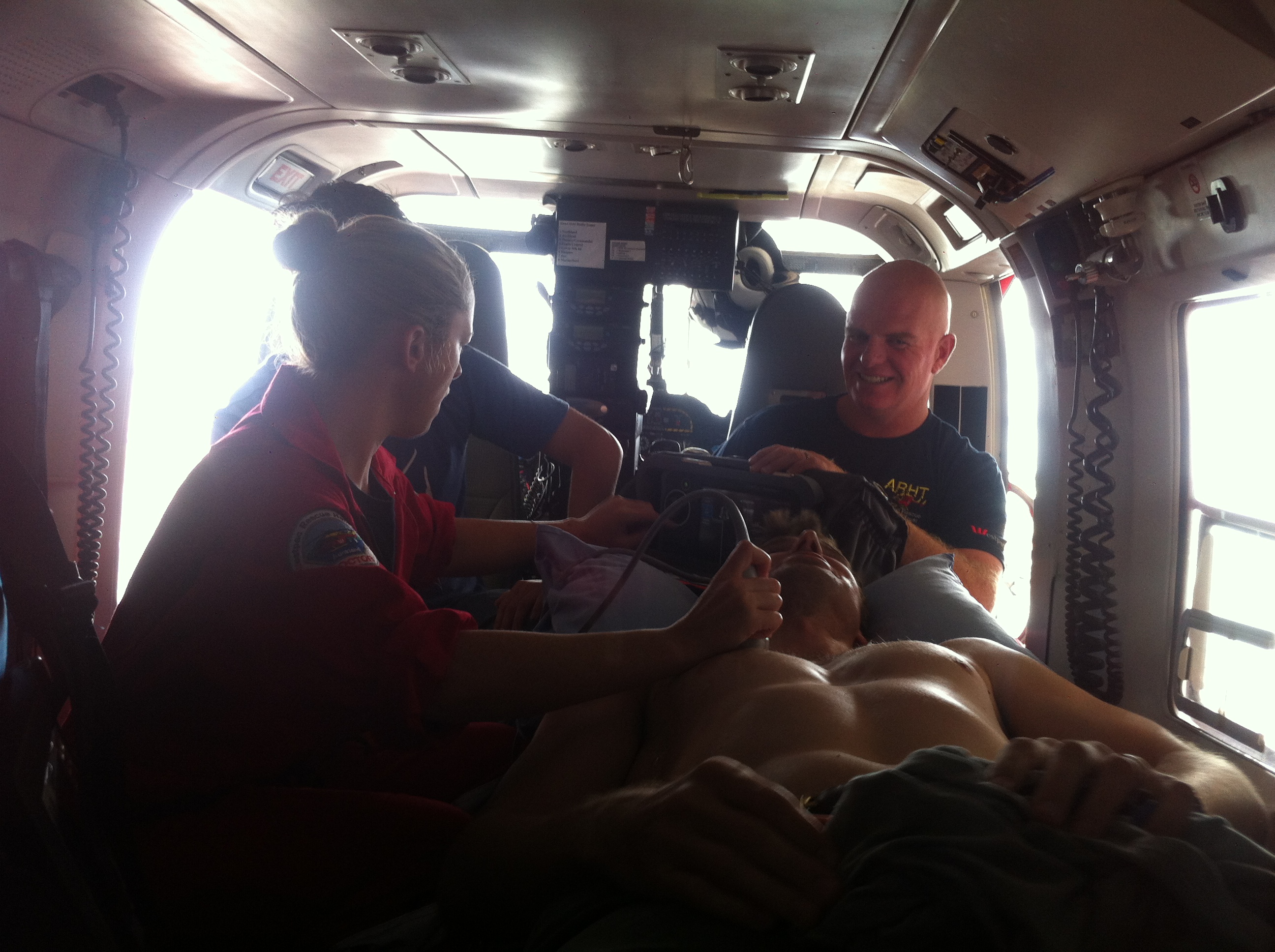
A little in-situ training. Enabled us to figure out optimal ergonomics and positioning for in-flight ultrasound.
In case you’re wondering, I donated my chest for this ultrasound to be done (free of charge!)
Summary
- Overall based on our evaluations of the process, it was a successful event with more case-based learning sessions planned
- Clinicians reluctant to intervene for pre-hospital pneumothorax unless unstable
- Strong communication among the team about the presence of a pneumothorax is essential and ultrasound greatly aids with this – affects both medical & operational decision making
- Ergonomics are important but dependent on each setting; however a standard approach in the machine might be appropriate for positioning of the ultrasound

Reblogged this on Sim and Choppers and commented:
Another successful case-based learning session at ARHT.