
An inadvertent leader in trauma research..!
There have been significant changes in the delivery of acute trauma care as a result of the military conflicts in Iraq and Afghanistan. One of the recent advances has been the advent of haemostatic dressings for haemorrhage control, which are now being used in the civilian as well as the military setting.
Auckland HEMS carries a product called Quikclot Gauze. This is an inert mixture of oxides of silica, sodium, magnesium, aluminium, and quartz. The compounds absorb water in a physical (not chemical) reaction, which concentrates platelets and clotting factors at the site of administration.
In 2008 the Journal of Trauma published a case series of the first 103 documented uses of Quikclot, including uses in military and civilian prehospital and hospital settings. The majority of uses involved extremity haemorrhage, often when direct pressure and tourniquets and direct pressure had failed. First responders found Quikclot to be 100% effective. Quikclot was ineffective in a handful of hospital cases, which involved moribund coagulopathic massively-injured patients. Heat generation from the physical reaction was an issue, with 3 patients sustaining burns, and a quarter of concious patients reporting ‘moderate to severe’ pain.
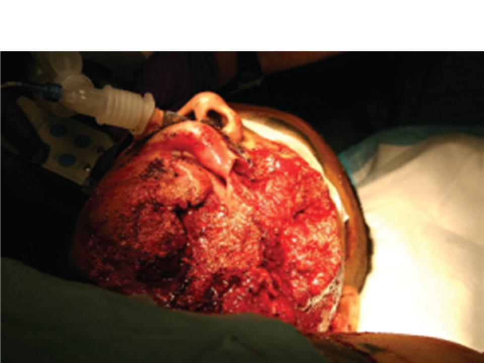
Wound from helicopter rotor blade, with Quikclot applied
There is also evidence from an animal model that Quikclot may allow sufficient haemostasis to reduce tourniquet time. This study involved a pig model of extremity haemorrhage and found that after haemostasis of a bleeding extremity had been acheived with a tourniquet and Quikclot, bleeding occurred only 20% of the time after tourniquet release, compared to a 100% failure rate with standard gauze dressings.
A systematic review of literature regarding haemostatic dressings was published in Injury in 2011, and can be found here. Overall data is scant and mostly observational/retrospective, but what is available suggests that haemostatic dressings like Quikclot should be a useful tool for controlling significant haemorrhage in our prehospital setting.
Full text pdfs for this post are here (secure area limited to ADHB staff only – ADHB has online subscription access to these journals through the Philson Library at the University of Auckland School of Medicine)