Prehospital Ultrasound – a new tool for our HEMS community

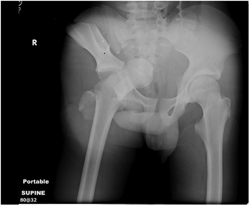
Several months ago, our HEMS service introduced a portable ultrasound machine onto our helicopters and so far it has been a great success!  While this blog post won’t be presenting the data we’re collecting, our physicians have reported it to be extremely useful. Most often we use it in the evaluation of a trauma patient to perform an eFAST (extended focused assessment with sonography in trauma) that includes assessment for free fluid in the abdomen but also importantly, lung ultrasound for the diagnosis of pneumothorax. Recently, I was part of a mission to transport a patient who had suffered a fall and there was question of a pneumothorax as reported by the ambulance team on scene. We were quickly able to perform an ultrasound of the lungs which ruled out pneumothorax. This enabled our pilot to fly at normal altitude rather than having to fly lower. Furthermore, as a clinician, it helped with decision making during transport as the patient still required treatment in hospital for other injuries. Knowledge that a pneumothorax was virtually unlikely allowed me to focus on other treatment priorities.

Picture of a similar model portable ultrasound that is being used at ARHT by HEMS physicians

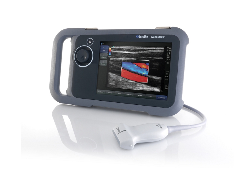
We’re using a similar model of portable ultrasound as pictured above at ARHT 

More recently, one of our physicians performed an ultrasound guided femoral nerve block to assist with pain management of a patient with a femur fracture. It worked brilliantly and the patient was transported with considerably less pain!

In the spirit of our new technology, I’ve reviewed what’s out there in the literature regarding prehospital ultrasound (and emphasis on HEMS). There’s very little but this is definitely a growing field!

A recent review of HEMS pre-hospital ultrasound feasibility was published with good results. They performed 144 pre-hospital scans. On average scans took less than 2 minutes with a symptom based approach to what region to scan. While there are some limitations in their methodology, they reported no false-positives compared with available clinical data which is important. In addition, overall sensitivity was 85% (though it should probably be reported for each indication). Nonetheless, this study adds support to the feasibility of prehospital HEMS ultrasound and documents what findings may be value in the field. In several cases, management was altered, for example when pneumothorax was diagnosed then chest drains were placed.

Another study just published, prospectively evaluated the utility of lung ultrasound in non-trauma patients with dyspnea in a pre-hospital setting. They used a focused approach (as pictured below) to specifically identify potential causes of dyspnea. In 68% of cases, physicians reported lung US as a useful tool.

Imaging sites for rapid assessment of lung using ultrasound in dyspneic patients in prehospital setting

Imaging sites for rapid assessment of lung using ultrasound in dyspneic patients in prehospital setting

They required physicians to complete the exam within 5 minutes as not to delay scene times. Pneumothorax was accurately ruled out in all cases, while a large pericardial effusion causing hemodynamic instability was properly diagnosed though it was only drained once in hospital. You might imagine however that if the patient deteriorated en route that emergent pericardiocentesis would probably be the next intervention so identification would be important.

Algorithm for evaluation of dyspneic patient in the prehospital setting with ultrasound

Algorithm for evaluation of dyspneic patient in the prehospital setting with ultrasound in conjunction with imaging sites of above picture. 

I’m not sure how to interpret their results when they reported that additional management approaches were taken in 25% of cases as a result of US. Primarily diuretics were administered after US given the diagnosis of pulmonary edema. In our setting, we don’t carry furosemide so this doesn’t directly apply though if perhaps properly delineating between pulmonary edema and COPD would be useful as nitroglycerin vs. nebulizers could be emphasized in subsequent therapy.

I believe that most of the benefit of prehospital ultrasound is in the injured patient however, as we see, there is growing evidence that it can be used similarly to how it’s used within the emergency department and ICU.

References

1.  Eur J Emerg Med. 2010 Oct;17(5):254-9. doi: 10.1097/MEJ.0b013e328336ae9e. Prehospital ultrasound in emergency medicine: incidence, feasibility, indications and diagnoses. Hoyer HX et al.

2. Eur J Emerg Med 2012 Jun;19(3):161-6. doi: 10.1097/MEJ.0b013e328349edcc. Prehospital chest emergency sonography trial in Germany: a prospective study. Neesse A et al.

Prehospital management of pelvic fractures

Will a pelvic binder help this?

Will a pelvic binder help this?

In the New Zealand trauma setting, blunt trauma is by far the most common mechanism. In the Auckland region it is not uncommon for us to manage patients with major pelvic trauma in ED (most commonly as a result of road trauma), and with the advent of the HEMS service our team are now dealing with this in the prehospital setting as well. The most notable case transported by the Auckland HEMS service in the last year involved a middle aged patient with a free-floating pubic symphysis (open bilaterally with actively bleeding groin wounds), a sacral fracture, and an ED arrival blood pressure of 66/40…)

This paper, published in 2007 by a UK trauma service, provides a nice overview of the prehospital management of pelvic trauma.

Take-home messages:

  • In patients who are obtunded (and therefore have an utterly unreliable clinical assessment) a pelvic fracture should be assumed to be present and a splinting device placed
  • in conscious patients, the presence of pelvic pain is a more reliable indicator of a fracture than palpation or compression of the pelvis
  • Reduction and stabilisation of pelvic fractures should occur as soon as possible after injury, while clotting mechanisms are still intact
  • Bleeding from pelvic fractures should be considered to be non-compressible, and therefore permissive hypotension (resuscitation to the presence of a radial pulse only) should be considered as a resuscitation strategy; NICE guidelines recommend 250mL boluses titrated to the radial pulse
  • There is a risk of patients becoming haemodynamically unstable following full log-rolls for spinal assessment (this has been reported in the ED setting); in the prehospital setting a roll to 15 degrees only will allow placement of a scoop
  • the handover to medical staff in ED should include advice not to remove the splint until a significant injury is excluded, including the fact that pelvic splints can provide excellent anatomical reduction leading to fractures potentially being missed and displacing once the splint is removed

The following is an instructional video showing the use of the SAM Sling, which is carried as standard kit on our helicopters:


Ever done an RSI in a helicopter? Here’s a recent simulation experience!

Recently at the base, we’ve been discussing the concept of improving our ergonomics and making our workspace (e.g. the helicopter) as functional as possible. We are continually looking to optimize our equipment to best serve our patients.  Any procedure in-flight will be considerably more difficult than if performed in a well controlled environment like the hospital so in-situ training within the helicopter is essential.

Today, Karl (one of our advanced paramedics) and I did some in-situ simulation of an RSI within the helicopter. We ran through a scenario with an unpredicted deterioration of a patient in flight that required an RSI. A review of the literature provides little guidance on the emergency airway management of patients while in-flight so approaches to such situations currently must be derived from simulation and retrospective reviews within your own program.

We discussed a few key concepts that should be considered as we move forward in pre-hospital airway management and overall care for acutely ill patients:

1. Patient positioning: ample evidence that patient’s should probably have some head elevation if possible during intubation (If you don’t believe me…check out this must read paper). This IS possible within the BK and it actually provided Karl with the best view when it was up near 40-45 degrees! Check out the following pics which demonstrates feasibility within the BK.

Patient is fully supine. Experts advocate "ear to sternal angle" but in our traditional position of supine you'll note that the ear is NOT at the sternal angle!

Patient is fully supine. Experts advocate “ear to sternal angle” but in our traditional position of supine you’ll note that the ear is NOT at the sternal angle!

And now, for a clear demonstration of “ear to sternal angle”. A position we should strive to do either to avert intubation or in preparation of an advanced airway.

A picture perfect view of the cords!

A picture perfect view of the cords! Patient at 40 degrees, and still able to intubate with a great view…even with the helmet on. Let’s integrate this!

2. Pack position: we decided that the airway/BMV pack would be removed from the Thomas pack and given to the intubating clinician immediately upon patient deterioration. This allowed the paramedic to have all necessary equipment for excellent airway management. The physician could then focus on drug administration and clinical decision making. We opened the Thomas pack fully beside the physician and placed the drug pack on the patient’s legs.

Note the drug pack on the patient's legs and the Thomas pack spread out to the right of the physician. This worked best in our setting.

Note the drug pack on the patient’s legs and the Thomas pack spread out to the right of the physician. This worked best in our setting.

Here’s what DIDN’T work.

This set up was very cumbersome if the drug pack is lying on a partially open Thomas pack. Another issue was the Thomas pack was still upright...and not lying flat.

This set up was very cumbersome if the drug pack is lying on a partially open Thomas pack. Another issue was the Thomas pack was still upright…and not lying flat. Also harder since we had to turn each time to get drugs rather than in front.

3. Apneic oxygenation: this is a bit trickier and something we’ll have to look at more closely to see what would be feasible since it will require 2 O2 sources. It was definitely challenging to get it set up when time constrained. (another must read paper on the value of apneic oxygenation).

Huge thanks to Karl for running through the sim case and providing value feedback on the ergonomics of the situation…what worked and what didn’t! We will all learn from this.