The importance of simulation in usability testing and hazard identification

photo-14

Auckland ED is conducting a lot of simulation training currently, with a recent focus on airway management.

Last week a teaching session was delivered by Sam Bendall (an Auckland HEMS doctor) on ‘intubation as a team sport’, which covered human factors in ED airway management and included the use of adjuncts like airway checklists. While Auckland HEMS has an RSI checklist, a similar tool has not been finalized for Auckland ED – this is under consideration currently. Following that teaching session, several airway checklists had nonetheless made their way into our resuscitation areas.

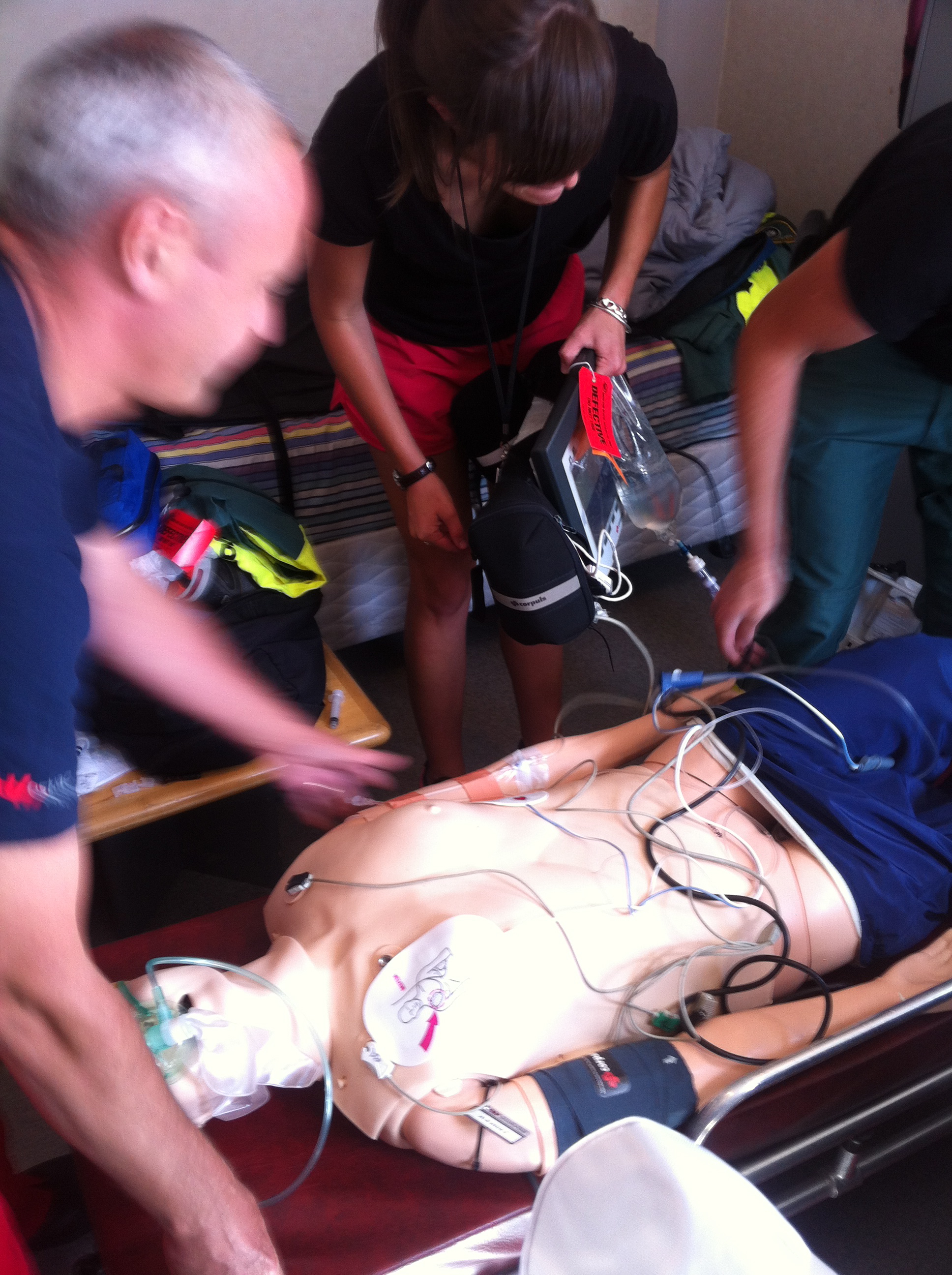
High-fidelity simulation training took place this afternoon, led by Sam, Mike Nicholls (another HEMS doctor), and Nancy Mitchell (Nurse Educator).

The first scenario involved a relatively junior team undertaking an emergent RSI. They performed admirably, and the outcome in practice would have been safe and successful. As an observer however, it was apparent that an airway checklist would have contributed to their confidence and comfort levels. (My personal opinion is that we should start using a checklist for ALL ED RSIs, independent of team seniority)

While watching the simulation I spotted a checklist taped to a whiteboard on a side wall. I assumed that was the airway checklist, and thought ‘that’s a clever position – it means the airway assistant can read out the checklist immediately prior to the RSI. They haven’t used it, I must bring this up at the debrief’.

At the end of the debrief, I inspected what I thought was the airway checklist, and found this:

HCA checklist

It wasn’t an airway checklist at all, but a restocking checklist!

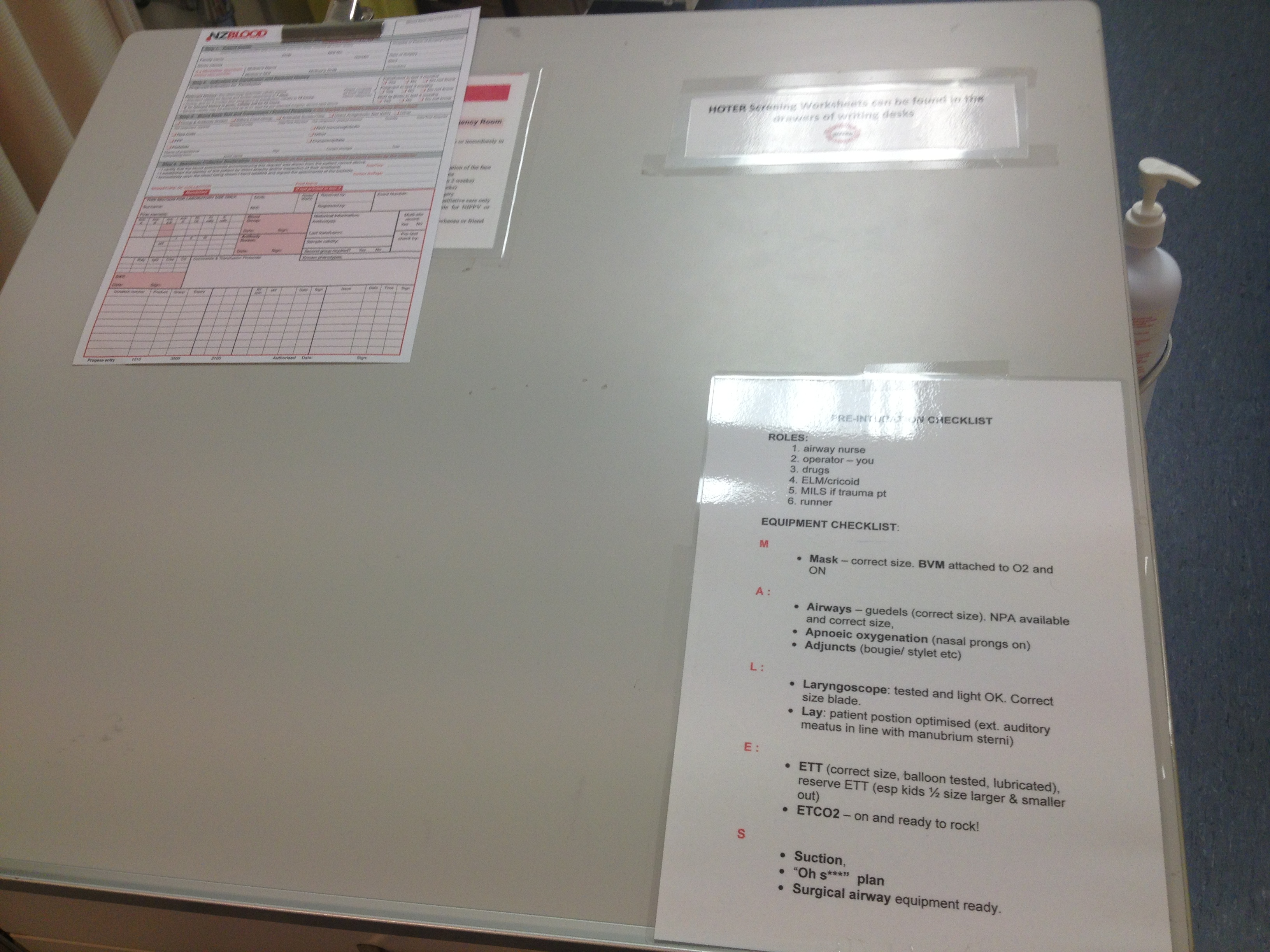
The actual airway checklist was here…

photo(3)

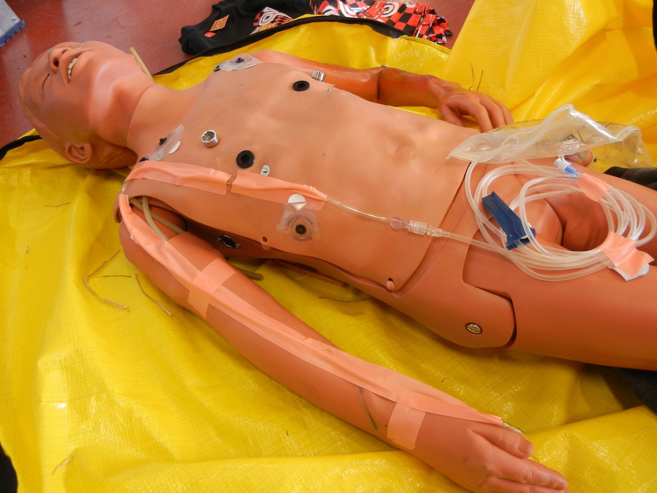
Tray following RSI (this is NOT how we set up!) Airway checklist bottom left

taped to the top of the airway trolley (logical) but unfortunately covered up by the airway tray, which is removed from the trolley and placed on top when setting up for airway management. This is a good example of a latent hazard.

I found a second copy of the checklist taped to the desk at the entrance to the resus bay – this is where the scribe (usually one of the senior nurses) stays during a resuscitation:

photo(2) copyAlthough this desk is frequently cluttered with paperwork during a resuscitation, accessing the checklist would be a simple matter, and would be done so by a senior person.

After looking at the positioning of these checklists, I came to two conclusions:

1) Simulation is a powerful tool for testing the usability of a new item or technique and identifying hazards

When considering logistics/ergonomics/equipment what actually happens in real life may differ from what we envisage mentally when we introduce something new. Simulation introduces stress, time-urgency, ergonomic elements and personnel elements that can rapidly reveal whether something new is going to be useful or not, or whether its introduction has inadvertently created hazards

2) We need to actively manage the environment we work in

When confronted with a critically ill patient, it is easy to focus on the scenario in front of us (there is usually quite enough to think about there!) and accept the physical environment as it is. By going a step further and ACTIVELY managing our environment to improve logistics and ergonomics we can increase our chances of a good outcome. This can occur both BEFORE we are confronted with a patient (eliminating the latent hazard above, for example) and DURING a resuscitation. This is particularly important in the pre-hospital setting, where both the relatively unforgiving helicopter and roadside environment provide a range of challenges not encountered in a resuscitation bay. As doctors I believe we have a lot to learn from our paramedic colleages in this area.

(NB – if someone becomes angry because that restocking checklist mysteriously disappears this week, I had NOTHING to do with it, nothing at all)

A medical student’s perspective at Auckland Rescue Helicopter Trust

For those of you who might have missed a few posts on here, you may not realize that HEMS has picked up a straggler along the way. I am a 5th-year medical student interested in pre-hospital and retrieval medicine, as well as anything high-stress and involving trauma and emergency. My role down at the base is to help out the HEMS doctors with setup of simulation scenarios, as well as keeping the simulation equipment and all our training packs stocked and ready to go. I am also keen to learn from everyone at the base on topics ranging from pre-hospital ultrasound to flying on instrument flight rules (IFR), as well as trying to convince myself and all the paramedics that one day I will be able to do a full-extension pull-up. Watch this space.

Today we unveiled our beautiful new manikins from Laerdal by using them in a simulated scenario on beta-blocker overdose.  We recruited the duty crew from the base, with the understanding that if a job came in they could easily leave the simulated scenario and go. We set it up as if the patient was in a remote medical centre after being retrieved by on-site paramedics from her home. The crew arrived to find a paramedic (yours truly) with the patient who had HR 30-35, bp 70/40, sats 97% on air, and RR 16/min. She had also had a 4-second period of asystole en route to the medical centre.

The team moving the patient onto the stretcher - ready for transport. Pads in place and ready for anything! Picture is a bit blurry because the team was moving with such efficiency & speed!

The team moving the patient onto the stretcher – ready for transport. Pads in place and ready for anything! Picture is a bit blurry because the team was moving with such efficiency & speed!

The remote control of the new manikin allowed us to simulate the heart rate and resps, whilst still maintaining fidelity of the scenario. This manikin also has the ability to moan, cough and respond yes or no, meaning the GCS could be fairly adequately ascertained. We used a sim technique we call ‘veining’ for cannula placement, which involves taping IV extension tubing up the arms of the manikin using skin-like tape with a cannula in the ACF, and then attaching an empty saline bag to the tubing to act as a reservoir. This allowed the participants to push drugs and run fluids, again preserving the fidelity of the scenario. We also used a piece of software called SimMon (for iPad), which can be set up to look like a regular monitor and will make appropriate noises when remotely controlled from iPhone (including that sweat-inducing desaturation beep). Our manikin can generate heart rhythms on our cardiac monitors but we use SimMon technology to supplement O2 saturation and blood pressure values.

The manikin with "veins" taped along the arms. It allows for actual IV starts and fluid administration. Everything is collected into the empty 1L NS bag.

The manikin with “veins” taped along the arms. It allows for actual IV starts and fluid administration. Everything is collected into the empty 1L NS bag.

This scenario utilized many things that I’m coming to realise are important in in-situ simulation. Firstly we used the duty crew, meaning that we didn’t have to get anyone to come in on their days off. But also that if a call came in for a job, our crew remains operational and can respond to that at any moment. So this makes our training highly efficient.   Secondly, this scenario was run on-site, including in the back of the chopper itself. Again excellent for fidelity and also for practicing techniques in a confined space. We are also vigilant about our labeling of training gear with bright red tags to ensure nothing from our training gear gets mixed with operational equipment.. A debrief time is equally as important as scenario-time, and this can be hard to facilitate if everyone disappears halfway through!

As a student, I found this scenario enlightening from both a simulation and medical point of view. I did some reading around beta-blocker overdoses and I found an excellent review about the use of high-dose insulin. Insulin has an increased inotropic effect on the heart, and clinical experience has shown that this has beneficial effects on patients who have overdosed on beta- and calcium-channel blockers. It’s also relatively cheap, readily available, and the dose to remember is easy – 1IU/kg bolus, then follow with 1-10IU/kg/hr infusion. Of course glucose needs to be monitored and a D5 infusion should be run whilst giving the insulin, and may need to be continued for up to 24h after the insulin has stopped. Monitoring the potassium is equally important, but remember the hypokalaemia is more due to cellular shift than overall potassium loss. There isn’t much data on the use of insulin in pre-hospital settings for such overdoses but it likely could be used during long flight times like inter-facility transfers. During our debrief, the clinicians felt it was more important to initiate early transport than high-dose insulin therapy in the field. But the discussion is pertinent and worth having.

All in all today was a good day for learning some key simulation techniques and some good emergency medicine. And apart from that, I managed to ask at least 14 irrelevant questions and steal 2 coffees, a yoghurt and half a banana from the lunchroom. So really a most successful morning.

Rossi Holloway

___________________

Full-text pdf for the reference above can be found here (secure area limited to ADHB staff only – ADHB has online subscription access to this journal via the Philson Library at the University of Auckland School Of Medicine)

Case Based Learning in the New Year: pneumothorax & lung ultrasound

Last week we ran another case-based learning session. The session consisted of a short discussion based around a case that we were tasked that involved a patient with a suspected pneumothorax.

We discussed the issues and challenges of managing a patient on the ground and in-flight with a pneumothorax. In addition, we discussed then practiced how we can use ultrasound as an added tool in the diagnosis of a pneumothorax in the prehospital setting.

To briefly summarize, I’ve divided up some discussion points

Medical

  • Both paramedics and doctors discussed the most important aspect in the patient with a pneumothorax in the pre-hospital setting was the clinical status
  • The ultrasound was noted to be extremely helpful for diagnosis however, presence of pneumothorax didn’t necessarily warrant intervention
  • Clinical condition was the overwhelming driver for intervention. The question arose regarding the role of ultrasound – “if the presence of pneumothorax did not necessarily mean intervention required, why use it?” In general, clinicians felt that knowledge about the condition would help make subsequent decisions in the case of deterioration
  • One theoretical approach was proposed – in a patient with pneumothorax that was reasonably stable, consider anesthesitizing & exposing the site for a chest drain then proceed with finger thoracostomy if deterioration. Several clinicians felt that it there was such concern to proceed with local anesthesia then probably a drain should just be placed.
  • In the patient with a left sided pneumothorax, there was strong agreement that loading the patient feet first such that the clinicians would have access to the left side (of our typically starboard loaded patient)
  • The likelihood of needle decompression success is only 50% – brief discussion about an anterior approach vs. a lateral approach

Operational

  • Knowledge regarding pneumothorax is key depending on the location of the patient. In situations on the east coast of the Coromandel then altitude becomes extremely important.
  • The early rule out diagnosis that the ultrasound can provide is very useful for managing flight plans
  • Weather was decided as a key factor that would alter management and it would impact possibly both medical decision making and flight operations
  • Placement of ultrasound in the machine: crewman/paramedic at the head of patient holding the machine with doctor on the patient’s right side
A little in-situ training. Enabled us to figure out optimal ergonomics and positioning for in-flight ultrasound. In case you're wondering, I donated my chest to science for this ultrasound to be done

A little in-situ training. Enabled us to figure out optimal ergonomics and positioning for in-flight ultrasound.
In case you’re wondering, I donated my chest for this ultrasound to be done (free of charge!)

Summary

  • Overall based on our evaluations of the process, it was a successful event with more case-based learning sessions planned
  • Clinicians reluctant to intervene for pre-hospital pneumothorax unless unstable
  • Strong communication among the team about the presence of a pneumothorax is essential and ultrasound greatly aids with this – affects both medical & operational decision making
  • Ergonomics are important but dependent on each setting; however a standard approach in the machine might be appropriate for positioning of the ultrasound


A simulation update: Latest session at the ARHT base

This week we ran an in-situ simulation with our duty crew (crewman, paramedic and doctor). We had great participation in a challenging scenario of massive hemorrhage in a blunt trauma patient.

As our simulation experience continues to grow we are always trialling new things. This past week we integrated several techniques that helped enhance the scenarios fidelity.

The scenario was a patient who had fallen off his motorbike at highspeed. There was a paramedic already on scene when our team arrived. The patient was in shock: BP 95/60, 130bpm, RR 28, 87% on room air, GCS 15.

Here’s a brief outline of what we did and why!

  • In-situ simulation: Make the most of the availability of your team. On the job training during a work day is a great way of maximizing educational opportunities. It doesn’t require that people come in on their day off and they still get paid while at work except their learning. We don’t use any expensive simulation centre – instead only using our training packs and equipment we were able to run this scenario at NO COST!
Mid way through a resus. We have all hands on deck, even getting our cameraman Matt to hold the IV!

Mid way through a resus. We have all hands on deck, even getting our cameraman Matt to hold the IV!

  • Set the scene with a video: using footage from the TV show Rescue 1 (filmed on our helicopters) we were able to begin the simulation with our team watching 2 minutes of a scenario to help them better picture the scene and envision the challenges of the local surroundings
  • Live patient actor: in scenarios that don’t require intubation this is especially powerful since we were able to capture our team’s ability to communicate with a live patient. Our patient had multiple traumatic injuries that was causing considerable pain. The team used managed the pain with ketamine and small doses of fentanyl. It was especially helpful to have a live patient since participants would receive real-time feedback if their pain regimen was working.
Having a live patient actor is a great asset and can add extra fidelity to the scenario. Definitely alters the way clinicians approach and speak with the patient.

Having a live patient actor is a great asset and can add extra fidelity to the scenario. Definitely alters the way clinicians approach and speak with the patient.

  • SimMon: I highly recommend this for anyone interested in doing in-situ simulation. Using an iPad and an iPhone, linked by Bluetooth (no Wifi needed) we are able to have a patient monitor with fully adjustable and modifiable vital signs! I have no relationship with the company that makes the app but we use it regularly and it’s must have for any educator running in-situ simulation. Available for download for less than $20NZD.
  • Ultrasound images for eFAST: Our doctor (Alana) performed a pre-hospital FAST and lung ultrasound. We had images and video downloaded ahead of time on a computer to show her the findings. This provided more realistic visual feedback that closely mimics a real clinical setting.
Alana checking out the eFAST findings on the laptop. Diagnosing pneumothorax & positive FAST

Alana checking out the eFAST findings on the laptop. Diagnosing pneumothorax & positive FAST

  • Integration of new medication: We are in the process of integrating a Tranexamic Acid protocol for trauma patients with suspected hemorrhage. This was our first time trialling the medication in a simulation setting. Great discussion around timing and especially helpful for our clinical team that we have clear guidelines when it can be administered.
  • Observation/Feedback by an industrial engineer: Tammy Bryan, is an industrial engineer from Auckland District Health Board, who joined us to observe our work with an interest in the ergonomics of scene set up. This was useful for a current state analysis and the beginning to work towards any changes that can make us more efficient!

Huge thanks to Bruce Kerr, Greg Brownson and Alana Harper who participated as our clinical and operational crew for the scenario. Also a huge thanks to Alice who was our live patient for the scenario. She did an outstanding job acting as a patient in pain with multiple injuries! Don’t worry, our team took care of her with lots of pain meds administered! And Chris was our paramedic who provided outstanding pre-hospital care before the team arrived