Inattentional Blindness – does this apply to pre-hospital medicine?

A recent study (not sure if it’s been published yet but will be soon) studied the ability of radiologists to accurately identify abnormalities on a CT scan. We’re talking board-certified, full fledged radiologists! I can’t take credit for coming across this paper – check out @TechnicalSkillz, ED physician in Toronto who tweeted the link. He has a real interest in cognitive biases and medical decision making. Anyways…I digress.

gorilla CT scan

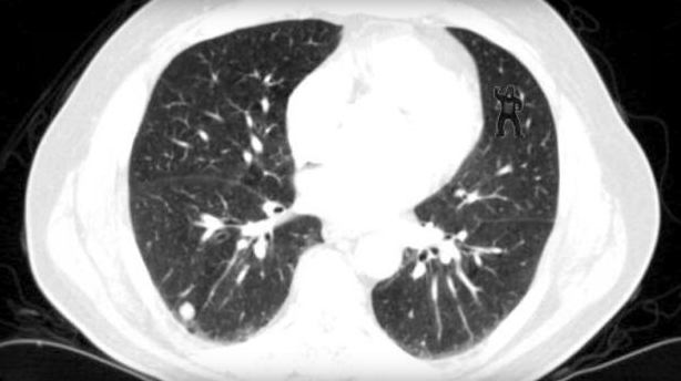
This image was presented to radiologists after they were told to look for abnormalities including lung nodules. Do you see the abnormality? Don’t worry…you don’t need to be an expert at reading CTs…it should be obvious!

24 radiologists examined this image and 20 were not able to identify the gorilla in the upper right corner! 20/24 didn’t see it! that’s unbelievable. They’re so focused on looking for other things that they glazed right over it. Using eye tracking technology, the reserachers were able to show that impressively 12 looked directly at the gorilla but still they didn’t see it!

This concept of inattentional blindness (or perceptual blindness) is

failure to notice an unexpected stimulus that is in one’s eyesight when other attention demanding tasks are being performed (Wikipedia)

I think this happens not infrequently in pre-hospital medicine and the emergency department. How many times does the patient turn out to have an entirely different presentation from what we hear over the radio call out or what’s written on the triage note. Right from the beginning we’re biased by the what we hear…it may be the local clinic calls with “a 73yr old with pneumonia”. Maybe there’s some shortness of breath to reaffirm this diagnosis but just as the radiologists were looking for nodules, we might be looking for pneumonia and miss the pneumothorax because we didn’t find out the patient fell earlier today. It could have been obvious if we just auscultated the lungs more closely but because we were thinking about crackles, we didn’t anticipate there would be absent breath sounds.

I think we’re especially vulnerable in the pre-hospital setting to inattentional blindness. We receive limited and often wrong information. Add that to time pressures to reduce scene times and it’s very easy to succumb to such cognitive biases.  We must implement cognitive checks to ensure that diagnoses are not missed. Standardized approaches to common presentations can help to ensure that critical diagnoses are considered regardless of the presentation. Efforts to ensure strong team communication will also enhance diagnostic abilities – maybe someone else on your team saw something you didn’t but failed to mention it.

Awareness of pitfalls around diagnostic errors must extend to pre-hospital clinicians. This will help us identify those gorillas! Check out some of the work by Pat Croskerry who’s a world leader in medical decision making, cognitive biases and diagnostic error (plus he’s Canadian, so he must be great!). Please note, the author of this post is Canadian which may be the reason for this conflict of interest!


Paediatric prehospital trauma care resources

whoops


With over half the Auckland HEMS team coming from a hospital that serves age 15 and up (and therefore not dealing with children on a daily basis), prehospital management of paediatric patients is a field to which we must pay considerable attention in our training. Simulation exercises have included paediatric scenarios on several occasions – our thanks to Mike Shepherd and Trish Wood from Starship Hospital for their assistance!

Below are some useful resources regarding prehospital care of paediatric trauma patients:

Podcasts from Dr Jeffrey Guy, Medical Director of PHTLS (right-click to download)

(he has also produced other PHTLS podcasts – found here)

A 2012 review of prehospital paediatric trauma from the Harbourview Medical Centre in Seattle can be found here. Take-home messages:

  • falls and MVA are the most common causes of paediatric trauma morbidity
  • despite a lot of research and intervention paediatric trauma patients are under-resuscitated on arrival compared to their adult counterparts
  • children have very different airways anatomically to adults; with full cervical spine immobilisation airway view can be improved with a towel under the shoulders to bring the neck into a neutral position
  • with a higher surface area/size ration children are more prone to hypothermia
  • due to their smaller blood volume a small amount of blood lost can represent a large percentage of their blood volume
  • contrary to traditional teaching, cuffed ETT are increasingly used at half a size smaller than the appropriate uncuffed ETT


Case Based Learning in the New Year: pneumothorax & lung ultrasound

Last week we ran another case-based learning session. The session consisted of a short discussion based around a case that we were tasked that involved a patient with a suspected pneumothorax.

We discussed the issues and challenges of managing a patient on the ground and in-flight with a pneumothorax. In addition, we discussed then practiced how we can use ultrasound as an added tool in the diagnosis of a pneumothorax in the prehospital setting.

To briefly summarize, I’ve divided up some discussion points

Medical

  • Both paramedics and doctors discussed the most important aspect in the patient with a pneumothorax in the pre-hospital setting was the clinical status
  • The ultrasound was noted to be extremely helpful for diagnosis however, presence of pneumothorax didn’t necessarily warrant intervention
  • Clinical condition was the overwhelming driver for intervention. The question arose regarding the role of ultrasound – “if the presence of pneumothorax did not necessarily mean intervention required, why use it?” In general, clinicians felt that knowledge about the condition would help make subsequent decisions in the case of deterioration
  • One theoretical approach was proposed – in a patient with pneumothorax that was reasonably stable, consider anesthesitizing & exposing the site for a chest drain then proceed with finger thoracostomy if deterioration. Several clinicians felt that it there was such concern to proceed with local anesthesia then probably a drain should just be placed.
  • In the patient with a left sided pneumothorax, there was strong agreement that loading the patient feet first such that the clinicians would have access to the left side (of our typically starboard loaded patient)
  • The likelihood of needle decompression success is only 50% – brief discussion about an anterior approach vs. a lateral approach

Operational

  • Knowledge regarding pneumothorax is key depending on the location of the patient. In situations on the east coast of the Coromandel then altitude becomes extremely important.
  • The early rule out diagnosis that the ultrasound can provide is very useful for managing flight plans
  • Weather was decided as a key factor that would alter management and it would impact possibly both medical decision making and flight operations
  • Placement of ultrasound in the machine: crewman/paramedic at the head of patient holding the machine with doctor on the patient’s right side
A little in-situ training. Enabled us to figure out optimal ergonomics and positioning for in-flight ultrasound. In case you're wondering, I donated my chest to science for this ultrasound to be done

A little in-situ training. Enabled us to figure out optimal ergonomics and positioning for in-flight ultrasound.
In case you’re wondering, I donated my chest for this ultrasound to be done (free of charge!)

Summary

  • Overall based on our evaluations of the process, it was a successful event with more case-based learning sessions planned
  • Clinicians reluctant to intervene for pre-hospital pneumothorax unless unstable
  • Strong communication among the team about the presence of a pneumothorax is essential and ultrasound greatly aids with this – affects both medical & operational decision making
  • Ergonomics are important but dependent on each setting; however a standard approach in the machine might be appropriate for positioning of the ultrasound